Category Archives: blog

Prostate Cancer

Prostate cancer in detail


Prostate cancer affects the prostate gland, the gland that produces some of the fluid in semen and plays a role in urine control in men.

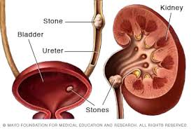
The prostate gland is located below the bladder and in front of the rectum. In the India, it is the Second most common cancer in men, but it is also treatable if found in the early stages.

In 2017, the American Cancer Society predicts that there will be around 161,360 new diagnoses of prostate cancer, and that around 26,730 fatalities will occur because of it.

Regular testing is crucial as the cancer needs to be diagnosed before metastasis.

Fast facts on prostate cancer:

Here are some key points about the prostate cancer. More detail is in the main article.

  • The prostate gland is part of the male reproductive system.
  • Prostate cancer is the most common cancer in men.
  • It is treatable if diagnosed early, before it spreads.
  • If symptoms appear, they include problems with urination.
  • Regular screening Is the best way to detect it in good time.

Symptoms

Prostate cancer is the most common cancer affecting men.
There are usually no symptoms during the early stages of prostate cancer. However, if symptoms do appear, they usually involve one or more of the following:

  • frequent urges to urinate, including at night
  • difficulty commencing and maintaining urination
  • blood in the urine
  • painful urination and, less commonly, ejaculation
  • difficulty achieving or maintaining an erection may be difficult

Advanced prostate cancer can involve the following symptoms:

  • bone pain, often in the spine, femur, pelvis, or ribs
  • bone fractures

If the cancer spreads to the spine and compresses the spinal cord, there may be:

  • leg weakness
  • urinary incontinence
  • fecal incontinence

Treatment

Treatment is different for early and advanced prostate cancers.

Early stage prostate cancer

If the cancer is small and localized, it is usually managed by one of the following treatments:

Watchful waiting or monitoring:

PSA blood levels are regularly checked, but there is no immediate action. The risk of side-effects sometimes outweighs the need for immediate treatment for this slow-developing cancer.

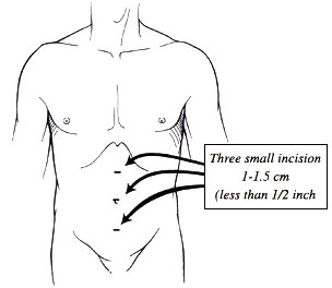
Radical prostatectomy:

The prostate is surgically removed. Traditional surgery requires a hospital stay of up to 10 days, with a recovery time of up to 3 months. Robotic keyhole surgery involves a shorter hospitalization and recovery period, but it can be more expensive. Patients should speak to their insurer about coverage.

Brachytherapy:

Radioactive seeds are implanted into the prostate to deliver targeted radiation treatment.
Conformal radiation therapy: Radiation beams are shaped so that the region where they overlap is as close to the same shape as the organ or region that requires treatment. This minimizes healthy tissue exposure to radiation.
Intensity modulated radiation therapy: Beams with variable intensity are used. This is an advanced form of conformal radiation therapy.

In the early stages, patients may receive radiation therapy combined with hormone therapy for 4 to 6 months.
Treatment recommendations depend on individual cases. The patient should discuss all available options with their urologist or oncologist.

Advanced prostate cancer

Advanced cancer is more aggressive and will have spread further throughout the body. Chemotherapy may be recommended, as it can kill cancer cells around the body. Androgen deprivation therapy (ADT), or androgen suppression therapy, is a hormone treatment that reduces the effect of androgen. Androgens are male hormones that can stimulate cancer growth. ADT can slow down and even stop cancer growth by reducing androgen levels. The patient will likely need long-term hormone therapy.

Even if the hormone therapy stops working after a while, there may be other options. Participation in clinical trials is one option that a patient may wish to discuss with the doctor.

Radical prostatectomy is not currently an option for advanced cases, as it does not treat the cancer that has spread to other parts of the body.

Fertility

As the prostate is directly involved with sexual reproduction, removing it affects semen production and fertility.
Radiation therapy affects the prostate tissue and often reduces the ability to father children. The sperm can be damaged and the semen insufficient for transporting sperm.
Non-surgical options, too, can severely inhibit a man’s reproductive capacity.
Options for preserving these functions can include donating to a sperm bank before surgery, or having sperm extracted directly from the testicles for artificial insemination into an egg. However, the success of these options is never guaranteed.
Patients with prostate cancer can speak to a fertility doctor if they still intend to father children.

What causes prostate cancer?

The prostate is a walnut-sized exocrine gland. This means that its fluids and secretions are intended for use outside of the body.
The prostate produces the fluid that nourishes and transports sperm on their journey to fuse with a female ovum, or egg, and produce human life. The prostate contracts and forces these fluids out during orgasm.
The protein excreted by the prostate, prostate-specific antigen (PSA), helps semen retain its liquid state. An excess of this protein in the blood is one of the first signs of prostate cancer.
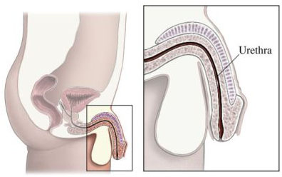
The urethra is tube through which sperm and urine exit the body. It also passes through the prostate.
As such, the prostate is also responsible for urine control. It can tighten and restrict the flow of urine through the urethra using thousands of tiny muscle fibers.

How does it start?

It usually starts in the glandular cells. This is known as adenocarcinoma. Tiny changes occur in the shape and size of the prostate gland cells, known as prostatic intraepithelial neoplasia (PIN). This tends to happen slowly and does not show symptoms until further into the progression.
Nearly 50 percent of all men over the age of 50 years have PIN. High-grade PIN is considered pre-cancerous, and it requires further investigation. Low-grade PIN is not a cause for concern.
Prostate cancer can be successfully treated if it is diagnosed before metastasis, but if it spreads, it is more dangerous. It most commonly spreads to the bones.

Stages

Staging takes into account the size and extent of the tumor and the scale of the metastasis (whether it has traveled to other organs and tissues).
At Stage 0, the tumor has neither spread from the prostate gland nor invaded deeply into it. At Stage 4, the cancer has spread to distant sites and organs.

Diagnosis

A doctor will carry out a physical examination and enquire about any ongoing medical history. If the patient has symptoms, or if a routine blood test shows abnormally high PSA levels, further examinations may be requested.
Tests may include:
• a digital rectal examination (DRE), in which a doctor will manually check for any abnormalities of the prostate with their finger
• a biomarker test checking the blood, urine, or body tissues of a person with cancer for chemicals unique to individuals with cancer
If these tests show abnormal results, further tests will include:
• a PCA3 test examining the urine for the PCA3 gene only found in prostate cancer cells
• a transrectal ultrasound scan providing imaging of the affected region using a probe that emits sounds
• a biopsy, or the removal of 12 to 14 small pieces of tissue from several areas of the prostate for examination under a microscope
These will help confirm the stage of the cancer, whether it has spread, and what treatment is appropriate.
To track any spread, or metastasis, doctors may use a bone, CT scan, or MRI scan.

Tips for Preventing Calcium Oxalate Kidney Stones

THE PATIENT EDUCATION piece “Tips for Preventing Calcium Oxalate Kidney Stones” was developed as a tool to instruct patients on the recommended nutrition treatment for nephrolithiasis. It is specific for calcium oxalate kidney stones.

Calcium and Oxalate

Oxalate binds to calcium in your stomach and intestines. When this happens, the oxalate won’t be able to get into your kidneys where it can form stones. Therefore, you should consume a moderate amount of calcium to bind the oxalate and help prevent kidney stone formation.

Include 2 or 3 servings of dairy foods every day. It is best to get your calcium from food rather than from supplements. If you do take calcium pills, take them with your meals.

Many foods contain oxalates, but only a few will result in high levels of oxalate in the urine. Avoid these high-oxalate foods:

Nuts and Nut Butters Tea (black and green) Rhubarb
Wheat Bran Chocolate Strawberries
Gelatin Spinach Black Raspberries
Beets and Beet Greens.


Drink More Fluid

You should drink at least 443.60ml of liquid each day.

This will dilute your urine and reduce the concentration of harmful substances that may cause kidney stones to form. At least half of the liquid you drink each day should be water.

Eat Less Salt/Sodium

Limit table salt to no more than ¼ teaspoon a day—measure it!

Omit salt in cooking; add ¼ teaspoon to fresh or prepared food to maximize flavor.

Only occasionally eat foods that are high in salt/sodium. These include the following:

Processed meats like bologna, hot dogs, and sausage; most fast foods; convenience foods; soup mixes, frozen soup, and canned soup; salted snack foods; boxed mixes for entrees or side dishes; high-sodium condiments such as pickles, olives, soy sauce, and steak sauce.

Limit Animal Protein

A high-protein diet can increase the oxalate level of your urine.

59ml serving is about the size of a deck of cards.

Vitamin C

Vitamin C may also be a problem because it can be converted to oxalate.

Do not consume more than 500 mg of vitamin C each day. This includes vitamin C from the foods you eat and from any oral supplement you may take.

Chronic Pelvic Pain

Chronic pelvic pain (CPD) is defined as a pain located in the pelvic area (lower abdomen) of more than 6 months. The painful sensation can be focused in different areas:

  • in the vagina
  • in the perineal area (between the anus and the vagina or between the anus and the scrotum)
  • in the scrotum
  • around the vaginal orifice

The fundamental problem of these patients is that very few urologist in West Delhi think about this syndrome when they come to the consultation. This makes the diagnosis take a long time, usually years, before starting appropriate treatment.

The causes of CPD syndrome are very numerous and varied: vaginal or pelvic surgical procedures, trauma (including repeated small injuries), inflammatory or infectious processes of the pelvis, etc. Usually, the pain is caused by irritation of the pudendal nerve or one of its branches. It is, therefore, what we call “neuropathic pain” against which the usual analgesics do nothing.

It is very characteristic that the pain is triggered or accentuated when the patient sits down and remits when lying down. In addition to pain, urinary symptoms may appear (urgency, frequency, incontinence, stinging, etc.); problems with the deposition; problems or difficulty having sex; erectile dysfunction; alterations of ejaculation, etc. All these associated symptoms are those that mislead the doctor who treats these patients, making the definitive diagnosis difficult.

The necessary study, in these patients, includes the following tests:

  • Physical exploration, locating those points that trigger the painful sensation (trigger points) and assessing the state of the pelvic musculature.
  • Magnetic Nuclear Resonance (NMR) of the pelvis, with special attention to areas where nerve entrapment could most likely occur
  • Neurophysiological study of the pudendal nerve

The PCD syndrome usually requires a long and multiple treatments, including:

  • Pharmacological treatment: As already mentioned, the usual analgesics are not effective in this situation. You must resort to antidepressant medications; state-of-the-art antiseptics; benzodiazepines; etc.
  • Infiltration of painful points with corticosteroids, local anesthetics, etc.
  • Physiotherapy treatments: electrostimulation, massages, etc.
  • Surgical treatment

Once the diagnosis is established, the therapies used are usually very effective and usually eliminate the pain and associated symptoms.

Kidney Stones in Children

Symptoms and diagnosis of kidney stones in children

Kidney stones are said to be able to hurt like childbirth, although at the moment, theirs is that births do not hurt. We are used to seeing them in adults, but in the pediatric age, they can also be seen. We talk about kidney stones. Next, we will reveal some of its characteristics.

Why kidney stones appear in children

Regarding its origin, there are calculations that appear spontaneously, others are secondary to abnormalities in the shape of the urinary tract, infections or metabolic disorders (among them, idiopathic hypercalciuria stands out, an entity that presents with an increase in the excretion of calcium in the urine). Obesity, sedentary lifestyle and heat (especially if the child does not adequately replenish the losses of fluids by sweating with water) favor their appearance.

Up to 50% of cases, there is a family component. That is, some component of the family has had some similar problem.

Symptoms, diagnosis, and treatment of kidney stones in childhood

From a clinical point of view, the most important manifestation is the pain. It is a very intense pain, located on both sides, and radiating to the abdomen. It is described as ‘exasperating’ (the child changes incessantly from a position), and associates with a vegetative procession of vomiting and cold sweat. The movement of the stone produces small erosions on the renal path, so the child bleeds. This bleeding is often evident in the urine.

The diagnosis can be established by the kidney hospital in Palam, West Delhi, and to document it we can resort to a urinalysis (in which we will see an increased number of red blood cells) and an imaging test (ultrasound, abdominal x-ray or tomography).

For kidney stone treatment, it is essential to hydrate the child (orally or intravenously, if he is very nauseous) and provide analgesia. In complex cases, before voluminous calculations, it is necessary to resort to surgery by the urologist in West Delhi. The calculations secondary to specific metabolic diseases require an individualized preventive kidney stone treatment in Uttam Nagar, West Delhi.

Direct Vision Internal Urethrotomy

Direct vision internal urethrotomy (DVIU) is surgery to repair a narrowed section of the urethra. This is referred to as a stricture. The urethra is the tube through which urine passes from the bladder to the outside of the body.

Reasons for Procedure

1. Urethral stricture is due to scarring of the urethra. This
2. Scarring may be caused by infection or injury. DVIU cuts
3. Through the scar tissue and
4. Opens the urethra.

Male Urethra

A urethral stricture can result in:
• Prostate problems in men
• Infections of the bladder or kidneys
• Inability to urinate or empty the bladder completely

Post-urethroplasty DVIU for isolated, recurrent strictures may be offered as a minimally invasive treatment option.

Mini percutaneous nephrolithotomy in the treatment of renal and upper ureteral stones

PCNL is a well-established treatment option for patients with large and complex renal calculi. In order to decrease morbidity associated with larger instruments like blood loss, postoperative pain and potential renal damage, a modification of the technique of standard PCNL has been developed. This is performed with a miniature endoscope via a small percutaneous tract (11–20 F) and was named as minimally invasive or mini-PCNL. This method was initially described as an alternative percutaneous approach to large renal stones in a pediatric patient population. Furthermore, it has become a treatment option for adults as well, and it is used as a treatment for calculi of various sizes and locations. However, the terminology has not been standardized yet, and the procedure lacks a clear definition. Nevertheless, mini-PCNL can achieve comparable stone-free rates to the conventional method, even for large stones. It is a safe procedure, and no major complications are reported. Although less invasiveness has not been clearly demonstrated so far, mini-PCNL is usually related to less blood loss and shorter hospital stay than the standard method.

In order to decrease morbidity associated with larger instruments like blood loss, postoperative pain, and potential renal damage, a modification of the technique of standard PCNL has been developed. This is performed with a miniature endoscope via a small percutaneous tract (11–20 F) and was named as minimally invasive PCNL or mini-PCNL or mini Perc.

CONCLUSION
Mini-PCNL was introduced as an alternative to the standard procedure in order to reduce morbidity associated with larger access tracts. Although less invasiveness has not been clearly demonstrated so far, mini-PCNL is related to less blood loss and shorter hospitalization. It is recommended for treatment of large renal stones in children and can be implemented in adults as well. Mini-PCNL seems to be a reasonable alternative for patients with a small-to-medium-sized stone, especially when a tubeless procedure is considered. Mini-PCNL is safe and is not related to serious complications.


Holmium Laser Enucleation of the Prostate (HoLEP)

Holmium Laser Enucleation of the prostate (HoLEP) is a modern alternative to the standard Transurethral Resection of the Prostate (TURP) procedure for bladder outflow obstruction due to BPH. It requires a short period of hospitalisation and an anaesthetic.

HoLEP can be performed on men of any age with urinary outflow obstruction caused by an enlarged prostate. It is particularly indicated in men with large prostates (over 60mls in size) and men on medications to thin the blood such as warfarin, aspirin or clopidogrel.

Advantages of HoLEP

  • There is no upper size limit of prostate that can be dis-obstructed – traditionally men with prostates over 100ml in size needed major open surgery
  • There is often less bleeding than after a TURP
  • Discharge is often quicker than after TURP at 1-2 days
  • The chance of recurrence requiring further surgery is very low
  • Unlike greenlight laser operations, large quantities of prostate tissue are sent for pathological analysis
  • The PSA generally drops to very low levels after HoLEP operations.

AIM OF HoLEP
The aim of HoLEP is to relieve pressure on the tube through which the urine drains (urethra) by anatomically enucleating the majority of excess benign prostate tissue. This is done under a general anesthetic with the help of a telescopic camera inserted through the penis. The three lobes of the prostate that are cored out intact are pushed into the bladder before being sucked up (morcellated) by a special instrument inserted through the telescopic camera. The pieces are sent for laboratory analysis just in case they might be found to be cancerous. A catheter is placed into the bladder to drain the urine while the raw surface heals, then left in place for around 24 hours before being removed on the day of discharge from the hospital. Sterile saline fluid is also irrigated into the bladder through the catheter to dilute any blood in the urine and prevent clots from forming.

Laparoscopic Pyeloplasty

Laparoscopic Pyeloplasty provides patients with a safe and effective way to perform reconstructive surgery of a narrowing or scarring where the ureter (the tube that drains urine from the kidney to the bladder) attaches to the kidney through a minimally invasive procedure. This operation is used to correct a blockage or narrowing of the ureter where it leaves the kidney. This abnormality is called a ureteropelvic junction (UPJ) obstruction which results in poor and sluggish drainage of urine from the kidney. UPJ obstruction can potentially cause abdominal and flank pain, stones, infection, high blood pressure and deterioration of kidney function. When compared to the conventional open surgical technique, laparoscopic pyeloplasty has resulted in significantly less post-operative pain, a shorter hospital stays, earlier return to work and daily activities, a more favorable cosmetic result and outcomes identical to that of the open procedure.

The Surgery
Laparoscopic pyeloplasty is performed under a general anesthetic. The typical length of the operation is 3-4 hours. The surgery is performed through 3 small (1cm) incisions made in the abdomen. A telescope and small instruments are inserted into the abdomen through these keyhole incisions, which allow the urologist in West Delhi to repair the blockage/narrowing without having to place his hands into the abdomen.

Ureteropelvic Junction (UPJ) Obstruction

A small plastic tube (called a ureteral stent) is left inside the ureter at the end of the procedure to bridge the pyeloplasty repair and help drain the kidney. This stent will remain in place for 4 weeks and is usually removed in the kidney hospital in West Delhi. A small drain will also be left exiting your flank to drain away any fluid around the kidney and pyeloplasty repair.

An Overview of Ureteroscopy and Flexible Uretero-Renoscopy

A special telescope is passed through the urethra, bladder, and into the ureter to the stone or to view the area of interest. The telescope may be rigid or flexible. The procedure is usually performed under general anaesthesia. A stone breaker, grasper or laser is passed up the ureteroscope to perform the procedure. This is a minimally invasive procedure that makes use of natural channels in the body; there are no cuts to the skin. It is a well tolerated and frequently performed procedure.

Ureteroscopy is performed for the following reasons:

  • Stones typically in the distal or middle ureter by rigid ureteroscopy that are unlikely to pass spontaneously or are causing significant discomfort.
  • Stones in the kidney that are not treatable by ESWL
  • To determine the reason for blood in the urine

Advantages of ureteroscopy

  • The stone is usually directly seen allowing the delivery of special instruments or lasers to break the stone. The ureteroscope is passed through natural channels in the body and involves no skin incisions. Providing the stone can be seen, there is a very high chance that the stone will be broken in one session.
  • Flexible ureteroscopy allows entry into all parts of the kidney so that all stones can be removed or vaporized provided they are of an appropriate size and accessible.

Success rates of ureteroscopy – The success rate of ureteroscopy is over 90% for the majority of stones that are treated this way. Success depends

  • Whether there are 1 or more stones present
  • How long the stone has been stuck
  • The location of the stone (wherein the kidney or ureter)
  • The size of the stone
  • Whether you have had previous surgery on the kidney
  • The experience of the urologist in Janakpuri treating you

Risks factors of ureteroscopy

  • Urine infection: this usually requires antibiotics only
  • Bleeding: this usually settles quickly
  • Damage to the ureter resulting in narrowing of the ureter (‘stricture’) or perforation: this is rare and may require stretching by a balloon and insertion of a JJ stent
  • Failure to break and retrieve the stone: an alternative procedure may be necessary
  • Perforation of the ureter: usually a JJ stent is required for a few weeks after such an injury
  • Detachment (‘avulsion’) of the ureter from kidney: this is very rare and is sometimes unavoidable, but may require open surgery to repair
  • Abdominal or back discomfort
  • Side-effects due to a JJ stent

Difference between rigid and flexible ureteroscopy

Rigid Ureteroscopy Flexible Ureteroscopy
Rigid ureteroscopy is performed literally with a rigid telescope. As such, it looks only in a straight line. Flexible ureteroscopy is performed with a very thin and flexible telescope that can perform almost a 180° turn and look back on itself. It is sometimes known as flexible ureterorenoscopy because it is possible to look into various parts of the inside of the kidney. Using a laser, stones can be vapourised and removed. Flexible ureterorenoscopy tends to be used for stones in the kidney and near the kidney in the upper ureter.
Rigid ureteroscopy is mainly used for stones in the lower and mid ureter closer to the bladder. Flexible ureter-renoscopy is more gentle than rigid ureteroscopy.

Precautions

  • Drink more fluid (especially 2 hours after meals and at night)
  • Adopt a diet appropriate to the type of stone. See calcium stone diet
  • Periodic X-rays or ultrasound to determine if more stones are being formed
  • Follow up in a stone clinic

Increase the Quality of your Life with Kidney Stone Treatment in West Delhi

In the modern lifestyle, we undergo a lot of changes in our dietary habits. Due to this kidney stones have become a common problem in people.  The number of people suffering from this problem is increasing day by day. Kidney stones are painful and result in decreased quality of life. A good step to prevent this is to take kidney stone treatment in West Delhi. It is important because once a stone is formed it will be risky for life.

Kidneystone treatment in West Delhi

The treatment of stones depends upon its type, severity of the condition and how long the person has suffered from its symptoms. In the case of small stones, there is no need for any treatment. The only thing that can be done in such cases is to wait for the stone to pass. Waiting for 4 – 6 weeks is safe only if the pain intolerable. Certain medications are also prescribed to manage the discomfort. Few types of surgical treatments are also available –

  • Shock Wave Lithotripsy (SWL) – In this procedure shock waves are targeted on the stones using ultrasound or X-rays that causes the stone to break into multiple small pieces. This results in passing of these small pieces through urine over a few weeks. This is a non-invasive treatment and the patient can go back to the home the same day of the procedure.
  • Ureteroscopy (URS) – This treatment involves passing of a ureteroscope up the ureter and into the kidney. By this way, urologist in Palam can remove the stone. If the stone is big and too large to remove in a single piece, it is broken down in several small pieces by using instruments.
  • Percutaneous Nephrolithotomy (PCNL) – It is carried out only when the stone is very large. It is carried out under general anesthesia and involves a half inch incision to be made. An instrument is passed through incisions to break the stone. The ability to suction fragments makes it the best treatment for large stones.

It is important for the people to come forward at an early stage of the stone problem to get best kidney stone treatment in West Delhi at an affordable cost. Early diagnosis and treatment will help you to stay healthy.